-
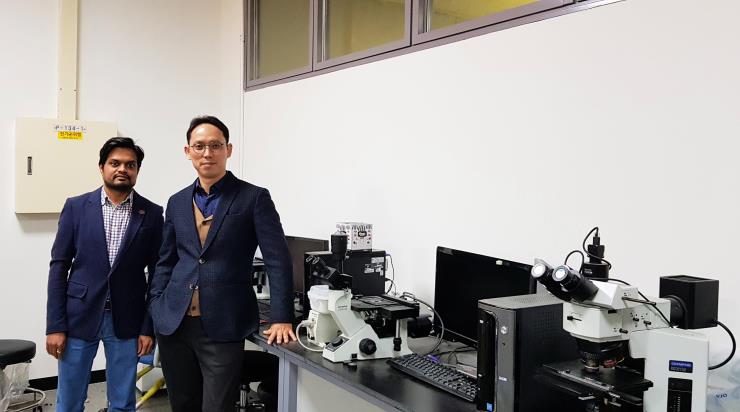
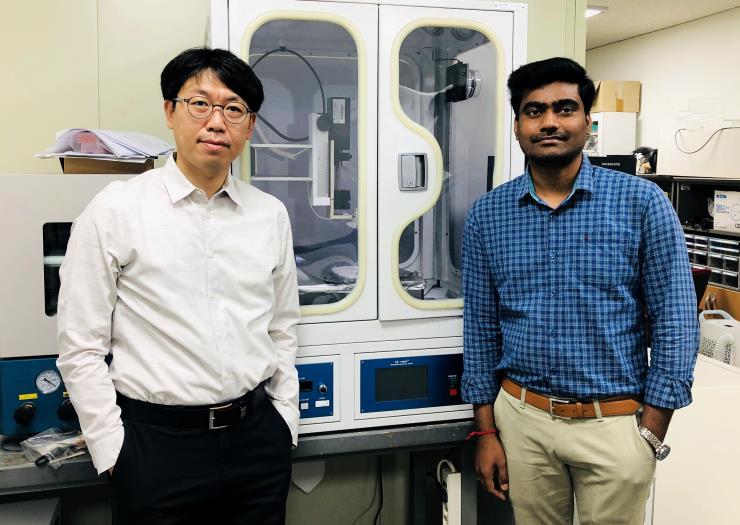
Professors Kim Wook and Yang Si-young’s team confirmed the osteoarthritis treatment mechanism of globular-shaped self-assembled hyaluronic acid nanoparticles. The recent finding is expected to contribute to improving in vivo safety and efficacy of linear hyaluronic acids used in chondroprotective agents for osteoarthritis. Professor Kim Wook (Department of Applied Chemistry & Biological Engineering, left in photo) and professor Yang Si-young (School of Medicine, right in photo) of Ajou University announced that the team has confirmed the chondroprotective efficacy of globular-shaped self-assembled hyaluronic acid nanoparticles and inflammation relief action. This means that target-specific drug carriers can become a potential therapeutic agent for osteoarthritis treatment.The study has been published in Biomaterials (IF 10.317), an international journal exploring the science of biomaterials and their translation towards clinical use, on June 19th with the title, “Self-Assembled Hyaluronic Acid Nanoparticles for Osteoarthritis Treatment.”The number of osteoarthritis patients continues to rise globally as lifespans have continued to lengthen, bringing to population aging. However, injection-type high-molecular-weight hyaluronic acids currently used in drugs and chondroprotective agents are not stable within a living body. The research team focused on self-assembled hyaluronic acid nanoparticles which are mainly used as in-vivo friendly, non-toxic, target-specific and long-acting drug carriers. As compressed globular-shaped self-assembled hyaluronic acid nanoparticles are relatively safer in vivo than linear hyaluronic acids, the team assumed that they are long-standing and provide increased inflammation relief. Accordingly, the team injected self-assembled hyaluronic acid nanoparticles into the knee joint of mouse models and observed through a multiphoton confocal microscope that the nanoparticles that penetrated the cartilage cell membrane conjugated with CD44 receptors and protected joint cartilage against osteoarthritis progression.This is evidence that multiple CD44-binding sites of nanoparticles conjugate with multiple CD44s at the same time, creating CD44 clustering in the cell membrane which effectively inhibits CD44 expression. The research team further explained that the drug carrier—self-assembled hyaluronic acid nanoparticles—itself, without any drug load inside, can inhibit CD44 receptors which aggravate joint cartilage erosion.In addition, the team reaffirmed that CD44 receptors can be a target for developing osteoarthritis treatments. CD44-deficient mouse models showed significantly lower osteoarthritis development compared to the control group with CD44 expression even when artificially inducing osteoarthritis in knee joints. The latest study suggests potential use of self-assembled hyaluronic acid nanoparticles as osteoarthritis treatments that interact with CD44. This recent finding is very meaningful as it provides a clue to overcoming the limitations of current linear hyaluronic acids and their low stability and inflammation-inducing effects due to product degradations.The study was sponsored by the Ministry of Science and ICT and the National Research Foundation of Korea as part of their Middle-grade Research Program and Bio/Medical Technology Development Program.<Self-Assembled Hyaluronic Acid Nanoparticles for Osteoarthritis Treatment Mechanism>
-
40
- 작성자OIA
- 작성일2021-08-03
- 8588
- 동영상동영상
-
A team of researchers led by Prof. Kim Sung-hwan, from the Departments of Physics and Energy Systems Research, has developed electronic skin capable of generating energy with a touch using silk protein nanofibers. This development is expected to be used as a next-generation bio-electronic material, generating electric energy or acting as sensory organs upon attachment to the human skin or soft robots. Prof. Kim announced successful development of electronic tattoos, which make it possible to draw circuits on silk protein nanofibers with carbon nanofiber ink using electrostatic-harvesting materials. Protein derived from silkworms is a skin-friendly, high-molecular biomaterial. The achievement was published online under the title, “Self-powered and Imperceptible Electronic Tattoos based on Silk Protein Nanofiber and Carbon Nanotubes for Human-machine Interfaces,” in Advanced Energy Materials (Impact Factor 25.245), a prestigious international journal covering energy materials, on May 11. Prof. Narendar Gogurla, Research Assistant Professor, participated as first author and worked together with Prof. Kim. There is a fast-growing body of research worldwide on materials for next-generation electronic healthcare devices for human-machine interfacing. Electrostatic-harvesting materials that leverage electrostaticity as an energy source are gaining more traction than ever. Electrostatic-harvesting materials can be used as a source of energy to move healthcare materials and artificial sensing organs that relay information related to human body movements. However, in order to increase energy efficiency, compatible materials should be used (e.g. hair and plastic show ideal electrostaticity), serving as an obstacle to generating electric energy in direct contact with human skin. In addition, the fact that the materials should be attached to human skin, no matter how uneven, while being composed of elements harmless to the human body, makes it harder to accelerate outcomes for research on electrostatic-harvesting materials. The team focused on silk proteins available from natural sources. Protein derived from silkworms is a skin-friendly, high-molecular biomaterial with superior physical and chemical properties. In the first step, the researchers make silk nanofiber paper, of a thickness equivalent to one-fiftieth of a strand of human hair, using electrospinning. Afterwards, they draw circuits on the silk nanofiber paper using carbon nanofiber ink and brush. Once the ink dries, the drawn circuits come out. Placing the circuits on slightly moist skin creates the electronic tattoo.The electronic tattoo is ultrathin, so it can be put on finely-wrinkled skin surfaces like finger pads while maintaining stability of its electric properties during everyday activities, besides taking showers. Electronic tattoos can easily be wiped off with wet wipes. Materials derived from silk nanofibers are harmless as the silk nanofibers are injected in the middle of the electronic tattoos.Interestingly, electronic tattoos attached to the human skin have the highest efficiency, signifying that other materials, including disposable gloves, should be worn to increase the efficiency of electrostatic energy. The harvested electric energy is sufficient to operate small electronic devices, such as LEDs and stopwatches. The fact that electric signals are generated with a single touch means that the tattoos can be used as artificial sensing organs. The team successfully drew pixel tattoos on the skin, proving that drawn circuits are capable of being converted into electric signals with the touch of a finger. Prof. Kim explained: “Although there has been significant progress in research on harvesting energy from the human body, the problems related to human-machine interfacing have been relatively neglected. Using proteins, of which natural skin is composed, interfaces capable of bridging the material difference between the human body and electronic devices can be developed.”He also added: “Our project is significant in that our invention can help increase the application of biomaterials to a variety of electronic devices. We hope to see our product applied to a wide range of healthcare products and soft robotics in the future.”The research was sponsored by the Rural Development Administration, the National Research Foundation of Korea, the BK21 FOUR, and the Gyeonggi-do Regional Research Center.Description: (Left) A conceptual diagram of a silk, electrostatic electronic tattoo, able to collect electric energy with a single touch on the skin using biocompatible electronic tattoos. (Right) An experiment on artificial sensing organs. Circuits drawn as pixel tattoos on the skin are capable of being converted into electric signals with the touch of a finger.
-
38
- 작성자OIA
- 작성일2021-08-03
- 7836
- 동영상동영상
-
A team of researchers led by Prof. Prof. Choi Sang-dun (from the Departments of Biological Sciences and Molecular Science & Technology) at Ajou University, announced the results of its study on drugs that are effective in preventing COVID-19. Prof. Choi (pictured), Prof. Kim Moon-suk (from the Departments of Applied Chemistry & Biological Engineering and Molecular Science & Technology), and S&K Therapeutics studied a number of anti-COVID-19 drugs approved by the US Food & Drug Administration and found that Remdesivir and Ledipasvir have potential to inhibit SARS-CoV-2 replication. S&K Therapeutics is a drug developer for autoimmune diseases, viral diseases, etc., and was established by Prof. Choi. The research findings were published in Cells, a prestigious international journal, under the title, “Remdesivir and Ledipasvir among the FDA-Approved Antiviral Drugs Have Potential to Inhibit SARS-CoV-2 Replication,” in April. Coronaviruses are positive-strand RNA viruses with a relatively larger virus genome. A genome is all genetic material of an organism, consisting of DNA or RNA. The coronavirus genome RNA is encapsulated by the nucleocapsid protein and polyadenylated with the spike glycoprotein on the viral envelope. The currently-developed COVID-19 vaccines especially target the spike glycoprotein.The team is currently developing materials that fight RNA-dependent RNA polymerase (RdRp). RdRp has a low probability of transformation, being the perfect target to control the reproduction of RNA viruses. The team, in an effort to respond in a timely manner to the rapid spread of COVID-19 and a surge in infections and deaths, focused on anti-coronavirus drugs approved by the FDA. Ajou University and S&K Therapeutics conducted an in silico screening of hundreds of antiviral drugs approved by the FDA through virtual screening first. In silico screening uses screening tools to make predictions about the behavior of different compounds. The team selected the first batch of drugs based on a series of simulations on anti-virus drugs and RdRp, and then identified the antiviral activities of SARS-CoV-2 in Vero E6 cells. As a result, Remdesivir and Ledipasvir were proven to be most effective. Vero E6 cells, derived from African green monkey kidney in 1962, have been used extensively for studies on viral infections. Vaccines against avian influenza, Rotavirus, polio, rabies, and many other diseases are produced with the use of Vero E6 cells. Prof. Choi explained: “The most pressing issue is that new coronavirus variants are being detected regularly. Thus, it is imperative that we develop a drug that prevents the proliferation of coronavirus over the long term.” He also added: “It is significant that we were able to discover that some anti-COVID 19 drugs can be used alone or with other medications.” Description: An assessment of the activities of SARS-CoV-2 antiviral drugs(a-c) The right Y-axis indicates cell viability while the left Y-axis indicates the activities of SARS-CoV-2 antiviral drugs.
-
36
- 작성자OIA
- 작성일2021-07-01
- 7789
- 동영상동영상
-
A team of researchers led by Prof. Seo Hyung-tak, from the Department of Materials Science and Engineering at Ajou University, has succeeded in developing water-splitting photocatalyst electrodes for hydrogen production. This breakthrough is expected to be useful in producing low-cost, high-efficiency photoelectrodes for hydrogen production in a non-polluting way. Prof. Seo Hyung-tak (pictured right) announced successful development of high-efficiency solar, water-splitting photocatalyst electrodes. The achievement was published online and entitled, “Enhanced solar water-splitting of an ideally doped and work function tuned {002} oriented one-dimensional WO3 with nanoscale surface charge mapping insights,” in Applied Catalysis B: Environmental (Impact Factor 16.683), an international journal, on May 6. Prof. Shankara S. Kalanur (pictured left), from the Department of Materials Science and Engineering, participated in the joint research as first author.The range of possible applications of hydrogen as an alternative source of energy for automobiles, electricity generation, and other industries has been expanding rapidly as of late. In order to produce hydrogen for such purposes, including fuel cells, reforming is the most common process. During the process, an amount of CO2 equivalent to nine times more than the hydrogen production weight is emitted. In an effort to reduce CO2 emissions, there is a fast-growing body of research worldwide to develop a solar, water-splitting technology using electricity and solar energy. Through water-splitting with the use of electric charges while inserting solar energy into semiconductor photocatalyst electrodes, CO2-free hydrogen is made. However, this process is a relatively low-efficiency form of production compared to existing fuel cell reforming methods. Therefore, improving the light reactions of photocatalyst electrodes, a major component in the solar water-splitting system, while securing long-term response durability is critical. Recently, photocatalyst electrodes have proven to be somewhat limited in achieving technological advancement; thus, a growing number of studies are now underway to develop tandem photocatalytic electrodes. However, the tandem structure makes the process complex, lowering the reproducibility of electrodes while triggering chemical instability between different materials. Under these circumstances, the team focused on tungsten trioxide (WO3) electrodes, which have been widely studied, but faced obstacles in terms of efficiency. Instead of using the two-component layered structure, Prof. Seo found that one-dimensional tungsten trioxide nanorods could be aligned with {002}, with high photocatalytic activities when a small amount (1.14%) of Yttrium (Y) is doped. To find optimal doping concentrations and processing, dozens of impurity concentrations were tested. Of particular note is that Yttrium(Y)-doped tungsten trioxide (WO3) showed an increase of up to 200% in photoelectric current, while the conversion efficiency of photoelectric currents into hydrogen was nearly 95 percent. Through a minimum amount of doping, various physical and chemical changes, including resistance reduction, changes in electronic structure, and changes in surface work function, were identified.Dr. Seo explained: “It is significant that even with the doping of minimum impurities into low-cost tungsten trioxide, nanostructured photocatalyst electrodes were made using high-efficiency, single-material elements. This made for highly-efficient conversion during the hydrogen production process. Our team is committed to ensuring its safety for commercial uses in the future.” The study was sponsored by the Research Support Program for New Researchers with Advanced Overseas Accomplishments, the Basic Research Support Program from the Ministry of Science and ICT and the National Research Foundation of Korea.
-
34
- 작성자OIA
- 작성일2021-07-01
- 7241
- 동영상동영상
-
Ajou University and Panoptics, a provider specialized in optics, have succeeded in developing multifunctional electronic tattoos using silk protein nanofibers. The invention is expected to be a next-generation healthcare device, enabling diagnosis and drug injection after attachment to uneven skin tissues after drawing the needed circuits on the skin. Prof. Kim Sung-hwan, Departments of Physics and Energy Systems Research (pictured left), announced successful development of electronic tattoos, making it possible to draw circuits on silk protein nanofibers with carbon nanofiber ink.The achievement was entitled, “Multifunctional and Ultrathin Electronic Tattoo for On-Skin Diagnostic and Therapeutic Applications,” and appeared on the Frontispiece page of the online edition of Advanced Materials, a prestigious international journal focused on materials science, on May 6. The research was conducted in collaboration with Panoptics. Primarily authored by Prof. Narendar Gogurla, Research Assistant Professor (pictured right), it was also written together with Mr. Kim Jang-sun, CEO of Panoptics, and three other authors. There is a fast-growing body of research worldwide on materials for next-generation electronic healthcare devices, as such devices are capable of detecting and analyzing vital signs automatically. Electronic materials that can provide as much flexibility and elasticity as actual human skin are needed and countless researchers have dedicated themselves to developing materials and devices that combine flexible substrates with electrodes and electronics to read and analyze vital signs from the human body. These materials are known as “electronic skin” today. Finding the right interface between electronic skin and actual human skin that can attach flawlessly to uneven skin surfaces, such as finger pads, and have enhanced biocompatibility is vital. Ultrathin electronic circuits using biocompatible materials have been very challenging to develop. Prof. Kim’s team sought to resolve this problem with proteins—particularly silk proteins available from natural sources. Protein derived from silkworms is a skin-friendly and high-molecular biomaterial with superior physical and chemical properties. In the first step, the researchers make silk nanofiber paper, with a thickness equivalent to one-fiftieth of a strand of human hair, using electrospinning. Afterwards, they draw circuits on the silk nanofiber paper using carbon nanofiber ink and brush. Once the ink dries, the drawn circuits come out. If the circuits are placed on slightly moist skin, this creates the electronic tattoos.These electronic tattoos are ultrathin and can be put on finely-wrinkled skin surfaces like finger pads while maintaining stability in their electric properties during our everyday activities, besides taking showers. Electronic tattoos are simple to wipe off with wet wipes. Materials derived from carbon nanofibers are harmless as the silk nanofibers are injected into the middle of the electronic tattoos.Electronic tattoos, as they are attached to the skin, can be used as electrodes for electrocardiography or electromyography. They can also be used in thermal therapy or drug injections. Weak electric currents transmitted to the electronic tattoos or exposing the tattoos to sunlight creates a certain level of heat through carbon nanofibers. The fact that light can be used to create heat means that electronic tattoos can be heated remotely, implying a broad and diverse scope of application. Together with the ability to inject drugs into silk proteins, controlling the temperature of the tattoos remotely will increase the efficiency of drug injections. Prof. Kim explained: “Although there has been significant progress in research on harvesting energy from the human body, the problems related to human-machine interfacing have been relatively neglected. Using proteins, of which natural skin is composed, interfaces capable of bridging the material difference between the human body and electronic devices can be developed.”He also added: “Our project is significant in that our invention can help increase the application of biomaterials to a variety of electronic devices. We hope to see our product applied to a wide range of healthcare products and soft robotics in the future.”Sponsored by the National Research Foundation of Korea, the BK21 FOUR, the Gyeonggi-do Regional Research Center (GRRC), and the Korea Institute of Energy Technology Evaluation and Planning, the electronic tattoo technology was developed in partnership with Panoptics. Patent applications in Korea and technology transfer have been approved as well.A conceptual image of silk protein-based electronic tattoos Circuits drawn with carbon nanofiber ink on silkworm-derived silk paper can be attached to the skin like a tattoo.
-
32
- 작성자OIA
- 작성일2021-07-01
- 7882
- 동영상동영상


